 |
 |
 |
 |
MORPHOLOGICAL CHANGES IN THE EPIDERMIS OF SUNFLOWER (Helianthus annuus L.) LEAVES INDUCED BY UV RADIATION
L.F. Hernández1,2 , L.I. Lindström1
and C.N. Pellegrini1,2
1 Departamento de Agronomía, Universidad Nacional
del Sur, Bahía Blanca (8000) and 2CIC, La Plata (1900),
Argentina. ( lhernan@criba.edu.ar
)
Introduction
UV radiation can affect growth, productivity and yield of different
crop plants. Changes in the morphology, anatomy and development of UV exposed
plants as well as the reduction of the leaf photosynthetic efficiency have
been reported (Teramura and Sullivan, 1994).
The sunflower shows tolerance to low doses of UV-B radiation (Barnes
et
al., 1990). Nevertheless alterations in its leaf morphology and reproductive
pattern in southern Argentina have been associated with recent increases
in the levels of UV-B (Sunflower Breeders, pers. comm.). Previous observations
using light microscopy (Hernández et al., 1999) revealed
some alterations at epidermal level and in the internal structure of leaves
(changes in the size and morphology of chloroplasts).
The objective of this work was to study the morphology of the leaf
surface of sunflower plants exposed to UV-A+B radiation using scanning
electron microscopy (SEM).
Material and Methods
Sunflower plants, cv. Dekasol 3881 DW were grown under controlled environmental conditions (18 h long-day photoperiod, 250 m mol s-1 m-2 PPFD at the canopy level and 26-24°C day-night temperature) in 2 L plastic pots containing garden soil. Plants were fertilized and periodically watered. They were exposed to UV- radiation (4 h day-1, 80 mmol.s-1m-) starting from 5 days from emergence (DFE) towards the end of floral differentiation (50 DFE). The source of UV were two 125W mercury lamps filtered with Pyrex (UV-A+B, 280-700 nm) or crystal (UV-A, 320-700 nm) following Sisson and Caldwell (1975). Leaf samples were taken after 65 DFE, fixed, processed for SEM (O'Brien and McCully, 1981), examined in a JEOL JSM-35CF scanning microscope at 10 kV and photographed.
Results and Discussion
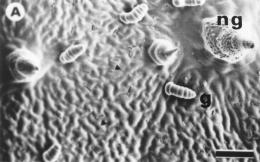
Control plants showed the typical epidermal structure described by many
authors (Seiler, 1997). In this particular genotype, two kind of trichomes
were identified: uniseriated pluricellular falcate non glandular and uniseriated
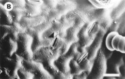
pluricellular non capitate glandular (Fig. 1A). Anomocytic stomatal complexes
and the absence of epicuticular wax deposits were also observed (Fig. 1B).
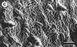
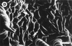
Treated plants differed from control ones showing higher density of glandular
trichomes and an evident collapse of the epidermal cells (Fig. 1C and D).
These results confirm the sensitivity of sunflower to the UV-A+B radiation
(Hernández et al., 1999). In spite of cellular damage at
surface level, the increment of pubescence observed in UV-treated plants
suggests an adaptive mechanism of tolerance to UV radiation (Fahn and Cutler,
1992).
References
Barnes, P.W., S. D. Flint and M. M. Caldwell. 1990. Amer. J. Bot.
77: 1354-1360.
Fahn, A. and D.F. Cutler. 1992. Xerophytes. G. Borntraeger. Berlin.
Hernández, L.F., L.I. Lindström and C.N. Pellegrini. 1999.
XVI International Botanical Congress, Saint Louis, Missouri.
O'Brien, T.P and M.E. McCully. 1981. The Study of Plant Structure.
Principles and Methods. Termacarphi Ltd., Melbourne.
Seiler, G.J. 1997. In: Sunflower Technology and Production. (A.A. Schneiter,
Ed.). Winconsin. pp: 67-111.
Sisson, W.B. and M.M. Caldwell. 1975. Photochem. Photobiol.,
21: 453-456.
Teramura, A. H. and J. H. Sullivan, 1994. Photos. Res. 39: 463-473.
|
|
|
|